91+ Animal Cell Parts Labeled
Especially made for those needing to remember them for exams etc. It is a double-membrane structure that surrounds the nucleus.

Basics Of Animal Cell Biology Lovetoknow
This game involves labelling parts of animal cells.

Animal cell parts labeled. Animal Cell Make sure that each part would have a definite function discussed. It is mainly composed of water salts and proteins. Find a picture and Label the parts of.
In eukaryotic cells the cytoplasm includes all of the material inside the cell and outside of the. The following animal cell diagram labeled show more parts of the cell. This organ controls the influx of nutrients and minerals in and out of the cell.
Sep 21 2018 - Printable animal cell diagram to help you learn the organelles in an animal cell in preparation for your test or quiz. It is also referred to as. Use the animal cell reference chart as a guide.
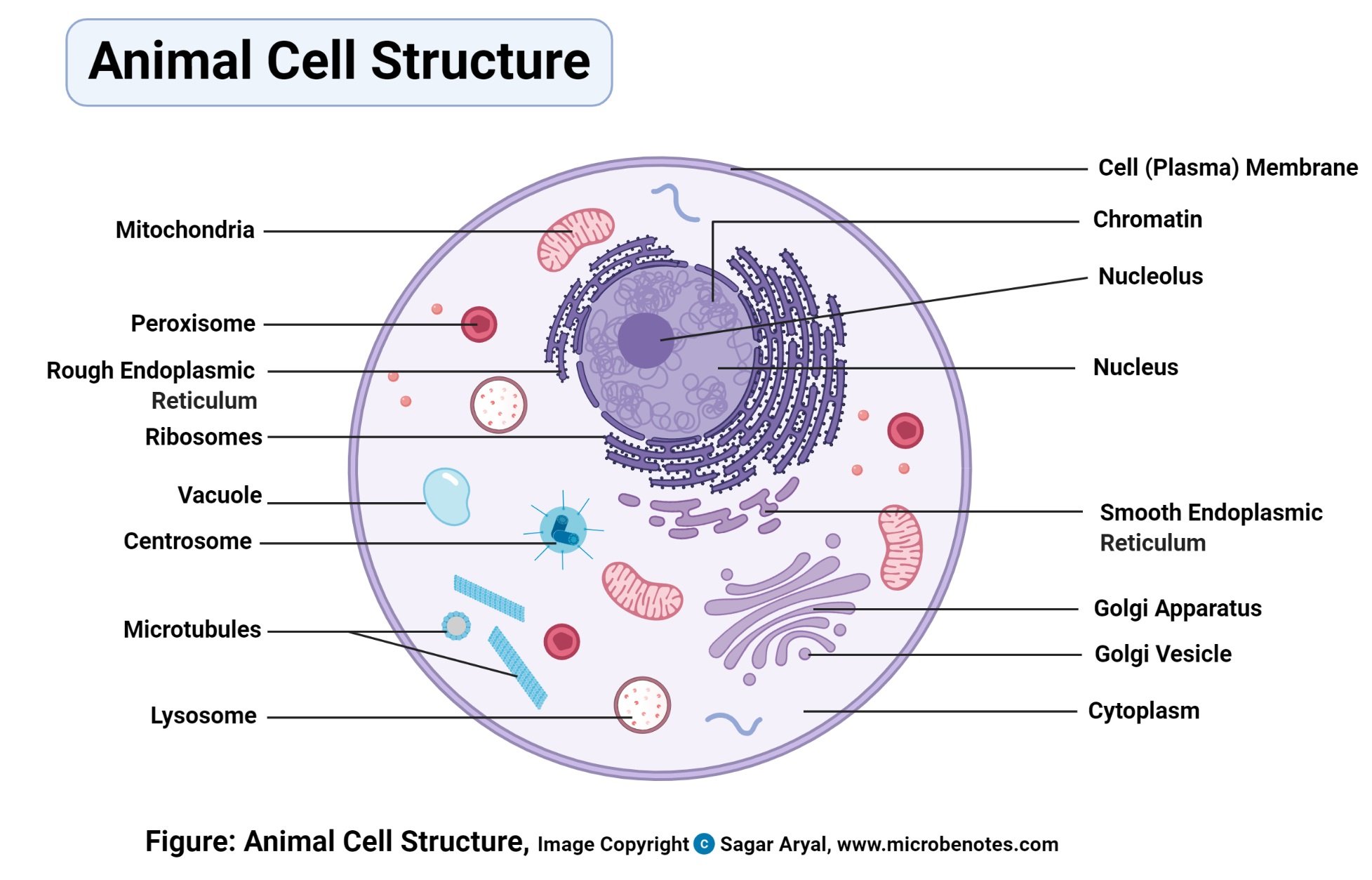
The cell membrane is the outermost part of the cell which encloses all the other cell organelles. Cytoplasm Jelly-like fluid that surrounds and protects the organelles. They are cell membrane nucleus nucleolus nuclear membrane cytoplasm endoplasmic reticulum Golgi apparatus ribosomes mitochondria centrioles vacuoles etc.
Its primary role is to. The membrane is selectively permeable and allows only certain molecules to pass through. Cell membrane surrounds the internal cell parts.
Animal Cell Parts Animal Cell Model Diagram Project Parts Structure Labeled Coloring and Plant Cell Organelles Cake. Learning about animal cells can be tricky at first so why dont we start you off relatively easy with this animal cell part labelling quiz. Animal cell diagram not labeled.
There are may parts inside a animal cell. In this one well be giving you a question referring to a given diagram and asking you to label it. Lets find out how many you get right.
4 An image of only the. Cell Membrane A double layer that supports and protects the cell. Animal Cell Structure Cell Membrane.
Controls passage of materials in and out of the cell cytoplasm everything inside of the cell membrane except for the nucleus light yellow nucleus control center of the cell. The cell membrane is a double-layered membrane made up of phospholipids that surrounds the entire cell. Animal Cell Picture with Labels Younger students can use the animal cell worksheets as coloring pages.
Labeled Animal Cell Diagram What Are the Various Parts of an Animal Cell. 0 005 Click on. Parts of an animal cell.
Plant cell Cytoplasm - is a thick solution that fills each cell and is enclosed by the cell membrane. Biology vector illustration Stock Vector and explore similar vectors at Adobe Stock. A thin semipermeable membrane layer of protein and fats surrounding the cell.
3 An image of the animal cell with only the nucleus in the center for reference. Nucleus The control center of the cell. Contains DNA light pink.
Your child will then use the first image to draw the rest of the parts of the cell and label each part. 2 An image of the animal cell with the 8 parts drawn and marked with arrows. Contains the DNA Nuclear Membrane Surrounds the nucleus.
Listed below are the Cell Organelles of an animal cell along with their functions. Allows materials in and out. Older students can be challenged to identify and label the animal cell parts.
Printable Animal Cell Diagram Labeled Unlabeled And Blank Finally an unlabeled version of the diagram is included at the bottom of the page in color and black and white. Create a spider map to identify and describe the different parts of the. Download The structure of an animal cell with labeled parts.
The child will then use the first image to copy the names of each part and eventually write them down by heart. Cytosol is the fluid present within a cell that is made up of water and ions such as potassium proteins and small molecules. Cells are made up of different parts.
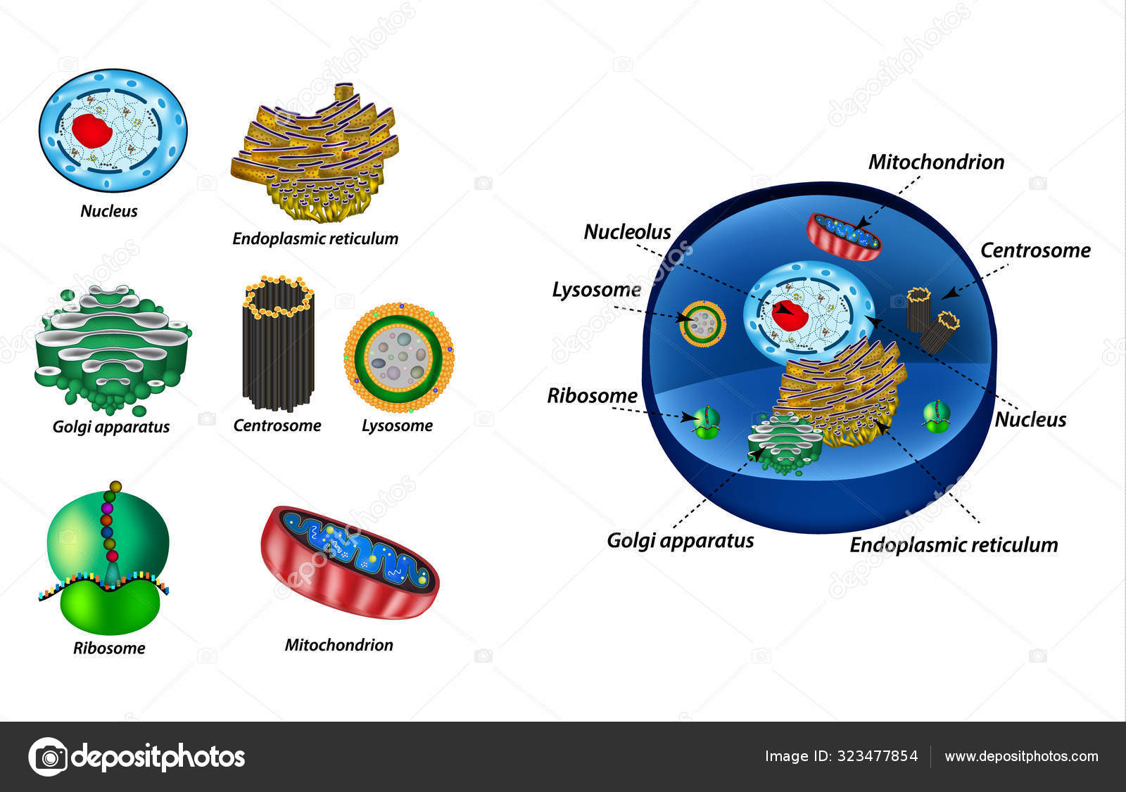
Centrosome Chromatin Cytoplasm Endoplasmic reticulum Golgi apparatus Lysosome Microtubule Mitochondria Nucleolus Nucleus Ribosome Vacuole 12 Create custom quiz. Lysosome Contains digestive enzymes that destroy damaged organelles and invaders. 5th grade science and biology.
21+ Animal Cell Under Microscope Labeled
Cells Blood Cell Nerve Cell Muscle Cell Skin Cell Ppt Video. Labels are a means of identifying a product or container through a piece of fabric paper metal or plastic film onto which information about them is.
B Why is it recommended to keep the stage of.
Animal cell under microscope labeled. Heres a diagram of a plant cell. Animal Cell Under Microscope Labeled. They are all typical elements of a cell.
You see that many features are in common. The information can be in the form of hand-written or printed text or. Its a thin slice.
So lets begin by drawing a rough-oval shape. Wide collections of all kinds of labels pictures online. We all do not forget that the human physique is quite problematic and a method I.
Labeled animal cell under electron microscope midbodyl. Animal cells under microscope animal cells under microscope labeled animal cells under microscope video animal cell under microscope 400x animal cell under microscope 40x animal cell under microscope 100x animal cell under microscope 10x animal cell. Day Ppt Video Online Download.
CZ08-001c Scalp - old skin flaking off hair. Photo Album By Darcy Plant And Animal Cells Under The Microscope. Illustrate Only A Plant Cell As Seen Under Electron Microscope.
CZ02-003p Stomach - cat parietal cells with. These are both specific types of. The tissue is composed of nerve cells neurons each of which is made up of a cell body and cell processes that carry impulses toward dendrites or away from axons the cell body.
Make your work easier by using a label. Labels are a means of identifying a product or container through a piece of fabric paper metal or plastic film onto which information about them is printed. The information can be in the form of hand-written or printed text or symbols and.
A Name the parts labeled A and B. Label Parts For Diagrammes Given Below And Identify Which One Is Plant Cell And Which One Is Animal Cell - Robert hooke was the first cytologist to identify cells under his microscope in 1665. Add a drop of purple stain specific for animals and cover with a cover slip.
The diagram is very clear and labeled. Diagram Of Animal Cell Under Electron Microscope. You should observe the cell membrane nucleus and cytoplasm Observation.
Wide collections of all kinds of labels pictures online. Wide collections of all kinds of labels pictures online. Draw a diagram of one cheek cell and label the parts.
The nuclei are stained with a red probe while the Golgi apparatus and microfilament actin network are stained green and blue respectively. You can observe this epithelial animal cell under microscope with high power. Observe the cheek cells under both low and high power of your microscope.
The cell membrane is the outer most part of the cell which. The following labeled drawings must be completed. Microscopic Animal Cells 82 images Microscopic Animal Cells.
Make your work easier by using a label. Labeled Animal Cell Under Electron Microscope. The figure below is a fine structure of a generalized animal cell as seen under an electron microscope.
See how a generalized structure of an animal cell and plant cell look with labeled diagrams. Diagrams from enchanted learning. Make your work easier by using a label.
CZ02-003d Stomach - cat parietal cells with. Although all animal cells look slightly different they will all be rounded without the sharp edges of plant cells and large enough to see at 100x under the microscope. Here is an electron micrograph of an animal cell with the labels superimposed.
Https Www Buffaloschools Org Cms Lib Ny01913551 Centricity Domain 1055 1 1 20intro 20to 20cells Pdf. Plant Cell Under Microscope Labeled. The outer edge of the cell.
CZ07-003c Skeletal Muscle - striated muscle. Module 5 Page 2. Labels are a means of identifying a product or container through a piece of fabric paper metal or plastic film onto which information about them is printed.
Diagram Of Animal Cell Under Electron Microscope Labeled. Sep 15 2014 - Learn the structure of animal cell and plant cell under light microscope. The animal cell is more fluid or elastic or malleable in structure.
Virtual Microscope Animal And Plant Cells Directions 1. B How is the structure labeled B adapted to its function. But at the same time it is interpretive.
In the following pages of this lab unit you will have an opportunity to examine a few of the many types of animal tissue. Illustrated in Figure 2 are a pair of fibroblast deer skin cells that have been labeled with fluorescent probes and photographed in the microscope to reveal their internal structure. Plant Microscopy Rosliston Astronomy Group Blog.
CZ01-008x Neurons - giant multi polar neurons. The granulated area is the cell Cytoplasm while the huge round part is the Nucleus. Monday April 5th 2021.
Within the cell there is a shape of round with a circular structure of granulated part on the epithelial cells. A Name the part of a light microscope used to bring an image of a specimen into sharp focus. Labeled animal cell under electron microscope 8745961 orig.
Cell is a tiny structure and functional unit of a living organism containing various parts known as organelles. The plant cell as more rigid and stiff walls.
81 Animal Cell Organelles
Ribosomes are small spherical organelles comprising 65 ribosomal RNA and 35 ribosomal proteins. There are 16 organelles in animal-like cells and 15 organelles in plant-like cells.

Cell Organelles Animal Biology Cell Cytoplasm Dna En Genetics Nucleolus Organelles Plant Glogster Edu Interactive Multimedia Posters
Ribosomes are very small organelles non-membranous and are present in thousands millions in some inside the cell.

Animal cell organelles. Each of these organelles performs a specific function for the cell. ATP fuels cellular processes by breaking its high-energy chemical bonds. 8th Jkbose The Cell Functions of organelles and similaritiesdifferences between Animal and Plant cell.
The cytoplasm is the fluid content inside the plasma membrane. It also contains many specialized cell organelles mitochondria golgi bodies ribosomes etc. Although both animal and plant kingdom falls under the eukaryotes multi-celled as opposed to prokaryotic which is single-celled animal cells have much more complex structure.
Animal cells contain ribosomes with four strands of RNA. Start studying Animal cells organelles. There are so many components that make up a cell and there is a huge difference between human animal and plantread more.
The brain or control center of the cell which directs all the cells activities. Terms in this set 14. Learn vocabulary terms and more with flashcards games and other study tools.
In this way what are the organelles of an animal cell and their functions. Ribosomes are not enclosed within a membrane but they are still commonly referred to as organelles in eukaryotic cells. The nuclear envelopenuclear membrane the chromatinchromosomes and the nucleolus.
As the site of cellular respiration mitochondria serve to transform molecules such as glucose into an energy molecule known as ATP adenosine triphosphate. The powerhouses of the cell mitochondria are oval-shaped organelles found in most eukaryotic cells. Therefore not every animal cell has all types of organelles but in general animal cells do contain most if not all of the following organelles.
Organelles in animal cells include the nucleus mitochondria endoplasmic reticulum Golgi apparatus vesicles and vacuoles. Structure of the plasma membrane opens a modal the cell membrane review opens a. Plant and animal cell.
Anatomy of an Animal Cell The study of cell theory helps us to understand each of the organelles in both plant and animal cells and what their jobs are. Some organelles that are found in animal cells but not in plant cells are. It is made up of 3 parts.
Organelles in animal cells include the nucleus mitochondria endoplasmic reticulum Golgi apparatus vesicles and vacuoles. It is the jelly-like substance present between the cell membrane and the nucleus.
24 Animal Cell Membrane Model
Using heavy metal staining Robertsons proposal also seemed to agree instantaneously with the Davson-Danielli model. Endoplasmic Reticulumsynthesizes carbohydrates and lipids.

Study Cell Structure Cell Membrane And Fluid Mosaic Model
Biology high school animal cell diagram.

Animal cell membrane model. If you chose to bake a nucleus cupcake youd also need to prepare a small amount of frosting dyed a third color. Nov 11 2014 - This is a selection of many project that were created by my seventh grade students during the 10 years that I taught at Slavens K-8 School in Denver Public Schools. In animals the plasma membrane is the outer boundary of the cell while in plants and prokaryotes it is usually covered by a cell wall.
Great animal cell model. It keeps the contents of the cell. As you refer to the animal cell model you will notice that this cell is lined by a double-layered cell membrane.
Legend opens a modal possible mastery. The model agreed with the thickness of cell membranes 4 nm estimated by H. Animal Cell Model Part II endoplasmic reticulum ribosome Golgi apparatus peroxisome and lysosomes.
Organelles have typed labels and students name and period are clearly labeled. The second color will be the cell membrane represented by the sides of the cake. Basically he suggested that all cellular membranes share a similar underlying structure the unit membrane.
Centrioleshelp to organize the assembly of microtubules. All pictures were taken by KR DuHoux. The following cell structures can also be found in a typical animal eukaryotic cell.
Animal cells differ from plant cells because the plant cell has a cell wall and chloroplasts and the animal. Jul 7 2013 teaching the cell can be fun and engaging for students. An animal cell is basically divided into two parts nucleus and cytoplasm.
Abby swan on January 16. Plant Cell Animal Cell Cell Membrane Plant Cell Vesicles Mitochondria Cell Wall Cell Membrane Nucleus Endoplasmi c Reticulum ER Nucleus Ribosomes Lysosomes Ribosomes Chloroplasts Cytoplasm Golgi Apparatus Mitochondria 16Do both cells in Model 3 have a nucleus. Model 3 Animal Cell vs.
The plasma membrane is commonly found in eukaryotic cells that contain membrane bound. By the 1950s cell biologists verified the existence of plasma membranes through the use of electron microscopy which accounted for higher resolutions. There were many more excellent projects that were not included primarily due to not having good photos.
The cell membrane is the substance that surrounds the entire cell. The nucleus is frequently called the brain of the cell. Jul 9 2012 - All living things are composed of cells which are one of two types.
Animal and plant cells are eukaryote cells. Styrofoam cardboard Tupperware empty milk container etc. Fricke in 1920-1930 by studying membrane capacitance.
This membrane not only separates the inner cell content from outside but also allows transportation of substances between the cell and surrounding. Great use of color. This is because it functions as the control center.
This lipidic bilayer model was the base for later adjustments and models of the cell membrane organization. The cell membrane is also known as the plasma membrane. Animal Cell Diagram Labeled Cakepins Com Animal Cell Project Animal Cells Model Animal Cell Model Project The.
Eukaryote cells have a nucleus whereas a prokaryote cell does not. Animal Cell Model Part III two types of temporary organelles involving in. The cell membrane is selectively permeable in nature consisting of a lipid bilayer with proteins glycolipids and cholesterol attached to them in a specific pattern.
See more ideas about cell model project cell model seventh grade. David Robertson used this method to propose the unit membrane model. 3D Animal Cell Project Model Parts of A 3D Animal Cell.
Cilia and Flagellaaid in cellular locomotion. Animal Cell Model Part I cell membrane cytosol nucleus and mitochondria. Cells prokaryotes and eukaryotes.
3d Cell Model Project Cells Project Animal Cell Cell Model Project Cell structure overview the major parts of the cell are the nucleus the control center. Eukaryote and prokaryote cells. For the cell membrane or cell wall.
I needed to make an animal cell model for my 7th grade project. Endoplasmic Ereticulum ER Golgi Apparatus Vacuoles Vesicles Yes. At this point you know that each eukaryotic cell has a plasma membrane cytoplasm a nucleus ribosomes mitochondria peroxisomes and in some vacuoles but there are some striking differences between animal.
All organelles are three-dimensional. The cell membrane is only one component of a cell. RETURN to CELL DIAGRAM.
This got me a 110.
59+ Animal Vs Plant Cell Organelles
The highest rank of biological. Animal cells dont have chloroplasts.

Which Organelles Exist In A Plant Cell But Not An Animal Cell Maxmirror
Lesson 2 Animal vs.

Animal vs plant cell organelles. Central vacuole cell wall and chloroplast. For example animal cells do not have a Cell Wall Chloroplasts or a Central Vacuole but plant cells do. A plant cell usually has a single large and prominent vacuole which is a bladder-like structure containing water and other solutes thus serving as a storehouse of the cell.
Both plant and animal cells are eukaryotic. They also have similar membranes such as cytoskeletal elements and cytosol. OVERVIEW OF CELL STRUCTURE VIDEO Both animal and plant cells.
Both plant and animal cells comprise membrane-bound organelles such as endoplasmic reticulum mitochondria the nucleus Golgi apparatus peroxisomes lysosomes. Chloroplasts and mitochondria have outer phospholipid bilayer membranes and circular DNA molecules like those of prokaryotic bacterial cells. This fascinating idea is called the Endosymbiont Theory or Endosymbiont Hypothesis for those who are more skeptical.
Below you can find a list will all of them animal cell organelles and their functions with and imagediagram to help you visualize where they are and how they look within the cell. Plant and animal cells. Here are some of the main difference between animal cells and plant cells along with all functions and organelles and what role each one plays within the cell.
Plant Cell vs Animal Cell. Animal cells are also the basic functional unit of life for animals constituting all cell organelles that perform a variety of functions to support the animals metabolisms. More often in replacement of lysosomes lytic vacuoles are found in plants performing the degradation.
A most important difference between plant cell and animal cell is that animal cell does not have a cell wall whereas plant cell does have a cell wall. Plant Cells CELL STRUCTURE Eukaryotes AnimalPlant Cells Remember we have 100 trillion cells that make up our body. Plant cells are basic functional units of plants constituting all cell organelles performing a variety of functions that support the plants metabolisms.
The most striking difference between animal cells and plant cells is that plant cells have three unique organelles. Plant cells have chloroplasts because they make their own food. Plant cells and animal cells both are eukaryotic cells that contain a nucleus and other organelles to perform necessary functions but the clear difference between them is the lack of cell wall vacuoles and chloroplast in the animal cell and the absence of flagella lysosomes and centrosomes in plant cells.
Similarities Differences with Chart Similarities Between Plant and Animal Cells. Plant and animal cells are both eukaryotic cells so they have several features in common such as the presence of a cell membrane and cell organelles like the. Lysosomes- Animal cells carry a larger amount of lysosomes organelle while plant cell also consists of them but the incidence is very rare.
Cell Organelles and Their Functions - Refer to pages 12 13 and fill in the following table for. Start studying Animal vs. Endoplasmic Reticulum Smooth and Rough Present.
It is very rare. Some biologists believe that mitochondria and chloroplasts within eukaryotic animal and plant cells may have originated from ancient symbiotic bacteria that were once captured by other cells in the distant geologic past. The plant cell can also be larger than the animal cell.
This membrane has about the consistency ofsalad oil. The animal cell has 13 different types of organelles ¹ with specialized functions. Chloroplasts are present in.
Learn vocabulary terms and more with flashcards games and other study tools. Centrosomes These are present in animal cells while being absent from the plant cells. The organelles unique for plant cells are vacuole cell wall and chloroplast shown in orange text.
Each cell of your body is encased in a tiny bubble of membrane. Round irregular shape Rectangular fixed shape Chloroplast. Animal cells may or may not contain one or many smaller vacuoles.
Various organelles are present in both animal cells and plant cells whereas some are present in either of them. Animal vs Plant Cells. Animal cells are mostly round and irregular in shape while plant cells have fixed rectangular shapes.
Below we are describing below what is found in all eukaryotic cells. ORGANELLES OF THE ANIMAL CELL AND THEIR FUNCTION. We summarize the major differences between plant and animal cells in this table.
Another difference between plant cells and animal cells is that animal cells can be all kinds of shapes and plant cells are usually boxy or rectangular. Synthesis of ribosomal RNA. Plant cells and cell organelles.
View Day-2-Plant-vs-Animal-Cell-Tablepdf from BIO 9900 at Australian Harbour International College.

